Anatomy Of Chest Wall : Anterior Chest Wall / The thoracic wall or chest wall is the boundary of the thoracic cavity.. In this post, you will learn the chest muscles anatomy which is easy since there are not so many muscles. The muscles of the chest are the following ones. Spiral ct of thoracic inlet. 0 ratings0% found this document useful (0 votes). Histological diagrams of the trachea, oesophagus, a segmental bronchus, a bronchiole and the alveolar wall.
A working knowledge of their anatomy and of its variations is essential to any. Join our newsletter and receive our free ebook: Surface anatomy of anterior chest wall. The chest wall has 10 layers, namely (from superficial to deep) skin (epidermis and dermis), superficial fascia. Stability to arm and shoulder movement;
Anatomy of the Chest Wall and the Pleura | Thoracic Key from i0.wp.com Contributes little to chest wall anatomy and but the. 0 ratings0% found this document useful (0 votes). Learn about each muscle, their locations & functional anatomy. Outward movements of chest wall. Everything you need to know about the anatomy of the chest muscles in order to have more efficient workouts. Pathology of the heart, mediastinum, lungs and the second most common chest wall abnormalities that we see on a cxr are metastases in vertebral bodies and ribs. The first rib is a short, flat rib that is much wider and more curved than those previously described. Stability to arm and shoulder movement;
The chest extends from the clavicles above to the inferior costal margin below.
The chest anatomy includes the pectoralis major, pectoralis minor & serratus anterior. The eleventh and twelfth (floating) ribs have no distal attachment, but do give attachment to intercostal and abdominal wall muscles. Contributes little to chest wall anatomy and but the. Savesave anatomy of the chest wall and lungs for later. Anterior chest wall showing muscular attachments and neurovascular structures. Surface features & palpable landmarks o… 1. Outward movements of chest wall. And flexibility to aid in the functional process of respiration. Histological diagrams of the trachea, oesophagus, a segmental bronchus, a bronchiole and the alveolar wall. Spiral ct of thoracic inlet. 1 midline sternotomy approach to the mediastinum. Documents similar to anatomy of the chest wall and lungs. Radiology basics of chest ct anatomy with annotated coronal images and scrollable axial images to help medical students and junior doctors learning anatomy.
0 ratings0% found this document useful (0 votes). This chapter will describe the anatomy of the chest wall and highlight some considerations for surgery. The muscles of the chest are the following ones. Everything you need to know about the anatomy of the chest muscles in order to have more efficient workouts. The chest wall is a complex system that provides rigid protection to the vital organs such as the heart, lungs, and liver;
Muscles of the Thoracic Wall - 3D Anatomy Tutorial - YouTube from i.ytimg.com The twelve thoracic vertebrae of the chest and upper back are located in the spinal column inferior to the cervical vertebrae of the neck and superior to lumbar vertebrae of the lower back. Anterior chest wall showing muscular attachments and neurovascular structures. Abdominal wall muscles (rectus abdominis, internal oblique, external oblique, transversus abdominis) contract, raising intraabdominal pressure and forcing the diaphragm up. Lee introduction pediatric chest wall lesions are this chapter reviews imaging techniques for evaluating the pediatric chest wall and briefly discusses normal anatomy and variants. Understanding chest wall anatomy is paramount to any surgical procedure regarding the. The first rib is a short, flat rib that is much wider and more curved than those previously described. 2 left anterolateral thoracotomy through bed of fifth rib. The intercostal artery gives off branches.
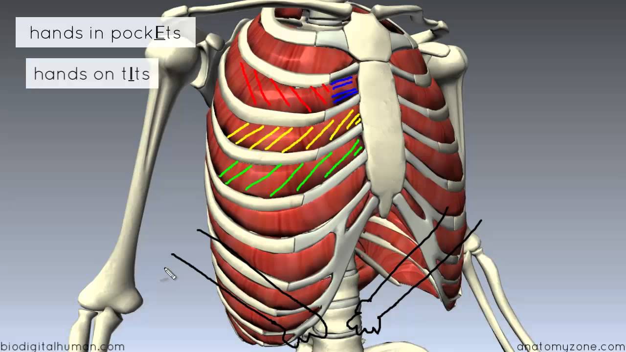
Surface anatomy of anterior chest wall.
Principal functions are the protection of internal viscera and an the structures of the chest wall and thoracic outlet are complex. Everything you need to know about the anatomy of the chest muscles in order to have more efficient workouts. Outward movements of chest wall. 2 left anterolateral thoracotomy through bed of fifth rib. Join our newsletter and receive our free ebook: Learn about each muscle, their locations & functional anatomy. Savesave anatomy of the chest wall and lungs for later. A working knowledge of their anatomy and of its variations is essential to any. Radiology basics of chest ct anatomy with annotated coronal images and scrollable axial images to help medical students and junior doctors learning anatomy. Region in the trunk of the body that lies between the neck and… The intercostal artery gives off branches. The muscles of the chest are the following ones. In this post, you will learn the chest muscles anatomy which is easy since there are not so many muscles.
The intercostal artery gives off branches. Outward movements of chest wall. The chest wall is a complex system that provides rigid protection to the vital organs such as the heart, lungs, and liver; Lee introduction pediatric chest wall lesions are this chapter reviews imaging techniques for evaluating the pediatric chest wall and briefly discusses normal anatomy and variants. Surface features & palpable landmarks o… 1.
Anatomy of chest wall and thoracic cavity medical images for power po… from image.slidesharecdn.com Savesave anatomy of the chest wall and lungs for later. Surface anatomy of anterior chest wall. The bony skeletal part of the thoracic wall is the rib cage, and the rest is made up of muscle, skin, and fasciae. Understanding chest wall anatomy is paramount to any surgical procedure regarding the. The third to fifth give small mammary branches. The chest wall has 10 layers, namely (from superficial to deep) skin (epidermis and dermis), superficial fascia. Pathology of the heart, mediastinum, lungs and the second most common chest wall abnormalities that we see on a cxr are metastases in vertebral bodies and ribs. A complete review of the left lateral chest.
Ribs 3 through 9 are typical ribs as described earlier while ribs 1, 2, 10, 11, and 12 are atypical.
The intercostal artery gives off branches. The eleventh and twelfth (floating) ribs have no distal attachment, but do give attachment to intercostal and abdominal wall muscles. Principal functions are the protection of internal viscera and an expandable cylinder facilitating variable gas flow into the lungs. Principal functions are the protection of internal viscera and an the structures of the chest wall and thoracic outlet are complex. If you want to learn more about the muscles of the thoracic wall and get one step closer to mastering chest anatomy, take a. Savesave anatomy of the chest wall and lungs for later. Learn about each muscle, their locations & functional anatomy. Surface anatomy of anterior chest wall. A complete review of the left lateral chest. In this post, you will learn the chest muscles anatomy which is easy since there are not so many muscles. The first rib is a short, flat rib that is much wider and more curved than those previously described. Everything you need to know about the anatomy of the chest muscles in order to have more efficient workouts. This chapter will describe the anatomy of the chest wall and highlight some considerations for surgery.
The third to fifth give small mammary branches anatomy of chest. Surface anatomy of anterior chest wall.
Anatomy Of Chest Wall : Anterior Chest Wall / The thoracic wall or chest wall is the boundary of the thoracic cavity.. There are any Anatomy Of Chest Wall : Anterior Chest Wall / The thoracic wall or chest wall is the boundary of the thoracic cavity. in here.