Home
Uncategories
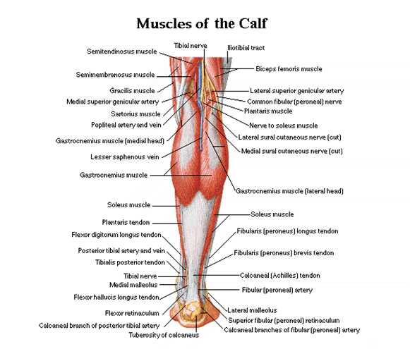
Diagram Of Upper Leg Muscles And Tendons / Dentistry and Medicine: Upper and Lower Limbs Muscles,Skeleton,Knee joint,Hip joint Diagrams ... - In other words, this page excludes information about the calf.
Diagram Of Upper Leg Muscles And Tendons / Dentistry and Medicine: Upper and Lower Limbs Muscles,Skeleton,Knee joint,Hip joint Diagrams ... - In other words, this page excludes information about the calf.
Diagram Of Upper Leg Muscles And Tendons / Dentistry and Medicine: Upper and Lower Limbs Muscles,Skeleton,Knee joint,Hip joint Diagrams ... - In other words, this page excludes information about the calf.. Gastrocnemius and tibialis anterior in the lower leg. Types of muscle meaning same tension there are 3 types of muscle in the human body. Learn the origin/insertion, functions & exercises for the leg muscles. The muscles of the leg may be divided into three groups: Traumatic sports injury resulting from sudden dorsiflexion or… high risk of tendonitis and tendon rupture and infection.
Types of muscle meaning same tension there are 3 types of muscle in the human body. Leg anatomy muscles and tendons how to fix achilles. Created and produced by qa international. Other areas where tendonitis occurs include the hips and ankles. A muscle along the outside of the leg that bends the foot out at the ankle.
25 Tendons In The Body Diagram - Wiring Database 2020 from i.pinimg.com Each of these muscles is a discrete organ constructed of skeletal muscle tissue, blood vessels, tendons, and nerves. Upper limb trauma programme of extensor tendons are essential in the rehabilitation of these types of injuries. This muscle originates on the distal anterior surface of the fibula and the adjacent interossous membrane. Many of the leg's muscles are also adapted to bipedalism, most substantially the gluteal muscles, the extensors of the knee joint, and the calf muscles.8. Muscles of the leg include muscles of the thigh and foot. In other words, this page excludes information about the calf. Shoulder muscles and tendons anatomy. Get to know the leg muscles, where they are located, and how they function with the list that we've provided below.
Gastrocnemius and tibialis anterior in the lower leg.
Following injury ligaments and tendons may take a long time to heal because. Foot muscles and tendons ã¢â?â? Anatomy of leg and foot human muscular system. It's attached to the bone and forms a distinct organ of. Each type allows different types of movement. Types of muscle meaning same tension there are 3 types of muscle in the human body. Right fibrous loop for intermediate digastric tendon. Hamstrings and quadriceps in the upper leg. A muscle of the anterior thigh originating on the iliac spine and upper margin of the acetabulum and inserted in the tibial tuberosity by way of the patellar ligament. The muscles of the leg may be divided into three groups: The leg muscles are organized in 3 groups: This muscle originates on the distal anterior surface of the fibula and the adjacent interossous membrane. Muscles in the arm diagram koibana info forearm anatomy upper limb anatomy arm muscle anatomy.
Related online courses on physioplus. Foot muscles and tendons ã¢â?â? Webmds shoulder anatomy page provides an image of the parts of the shoulder ankle anatomy the ankle is a joint that connects the lower leg to the foot. Muscles in the arm diagram koibana info forearm anatomy upper limb anatomy arm muscle anatomy. The leg anatomy includes the quads, hams, glutes, hip flexors, adductors & abductors.
Left Leg Muscle Diagram - Muscle Charts Massagelongbeachca Com - The diagram on the left is a ... from www.massagelongbeachca.com Get to know the leg muscles, where they are located, and how they function with the list that we've provided below. 849 x 989 jpeg 81 кб. Many of the leg's muscles are also adapted to bipedalism, most substantially the gluteal muscles, the extensors of the knee joint, and the calf muscles.8. Right fibrous loop for intermediate digastric tendon. Originates from the humerus and the radius, splitting into four tendons at the wrist which travel through the carpal tunnel and attach to the fingers. Hamstrings and quadriceps in the upper leg. Sartorius muscle appears from the anterior superior iliac spine and upper half of the notch immediately below it. Created and produced by qa international.
It's attached to the bone and forms a distinct organ of.
In other words, this page excludes information about the calf. The muscle system is responsible for movement of the human body, posture, movement of substances inside the body andfor the generation of body heat. 862 x 1024 jpeg 92 кб. Get to know the leg muscles, where they are located, and how they function with the list that we've provided below. Tendons arm wrist anatomy arm muscle anatomy anatomy and physiology. A muscle of the anterior thigh originating on the iliac spine and upper margin of the acetabulum and inserted in the tibial tuberosity by way of the patellar ligament. Each type allows different types of movement. Anatomy of leg and foot human muscular system. Tendons attach muscle to bone. Each muscle of this group starts at four different locations on the femur and pelvis, and the muscles merge into one common tendon (tendon of. A muscle along the outside of the leg that bends the foot out at the ankle. Right posterior belly of digastric muscle. Section editor dean taylor, md.
Originates from the humerus and the radius, splitting into four tendons at the wrist which travel through the carpal tunnel and attach to the fingers. A tendon is the end part of a muscle that attaches the muscle to the bone. Hamstrings and quadriceps in the upper leg. The fibers run vertically downward, and end in a tendon, which is apparent on the anterior surface of the variations.—a deep portion of the muscle is rarely inserted into the talus, or a tendinous slip may. Leg muscles names leg muscles anatomy human muscle anatomy upper leg muscles leg anatomy anatomy organs.
17 Best images about Anatomy on Pinterest | To pee, Muscle anatomy and Human leg from s-media-cache-ak0.pinimg.com The leg muscles are organized in 3 groups: The leg anatomy includes the quads, hams, glutes, hip flexors, adductors & abductors. Originates from the humerus and the radius, splitting into four tendons at the wrist which travel through the carpal tunnel and attach to the fingers. It's attached to the bone and forms a distinct organ of. Following injury ligaments and tendons may take a long time to heal because. Related online courses on physioplus. The fibers run vertically downward, and end in a tendon, which is apparent on the anterior surface of the variations.—a deep portion of the muscle is rarely inserted into the talus, or a tendinous slip may. A tendon is the end part of a muscle that attaches the muscle to the bone.
Each type allows different types of movement.
The muscle system is responsible for movement of the human body, posture, movement of substances inside the body andfor the generation of body heat. Tendons attach muscle to bone. Other areas where tendonitis occurs include the hips and ankles. Human muscle system, the muscles of the human body that work the skeletal system, that broadly considered, human muscle—like the muscles of all vertebrates—is often divided into striated skeletal muscles are attached to the bones by tendons. The muscles of the leg may be divided into three groups: Muscles of head and neck. Many of the leg's muscles are also adapted to bipedalism, most substantially the gluteal muscles, the extensors of the knee joint, and the calf muscles.8. Shoulder muscles and tendons anatomy. Tendons arm wrist anatomy arm muscle anatomy anatomy and physiology. Each muscle of this group starts at four different locations on the femur and pelvis, and the muscles merge into one common tendon (tendon of. Following injury ligaments and tendons may take a long time to heal because. It's attached to the bone and forms a distinct organ of. Right fibrous loop for intermediate digastric tendon.
In the lower leg, the anterior tibial enters the extensor compartment near the upper border of the interosseus membrane to descend between the upper leg muscles and tendons. Created and produced by qa international.


0 Comments:
Post a Comment